Age-related macular degeneration is the most common cause of severe loss of eyesight among people 50 and older. Only the centre of vision is affected by this disease. It is important to realise that people rarely go blind from it.
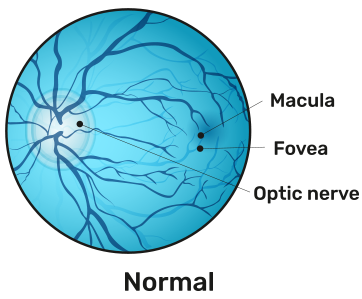
AMD affects the central vision and with it, the ability to see fine details. In AMD, a part of the retina called the macula is damaged. In advanced stages, people lose their ability to drive, to see faces, and to read smaller print. In its early stages, AMD may have no signs or symptoms, so people may not suspect they have it.
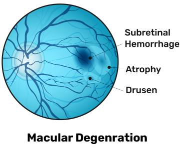
The presence of drusen, which are tiny yellow deposits in the retina, is one of the most common early signs of age-related macular degeneration. It may mean the eye is at risk for developing more severe age-related macular degeneration. These will be visible to your doctor during an eye exam.
Types and Causes
The two primary types of age-related macular degeneration have different causes:
Dry
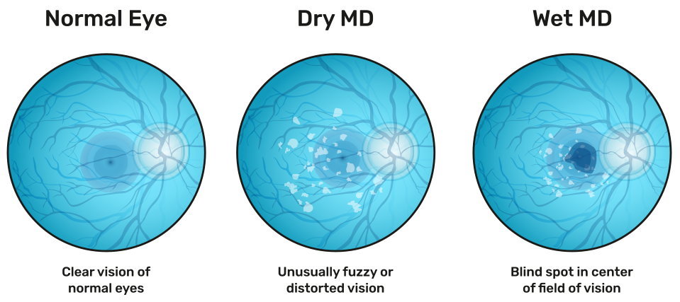
This type is the most common. About 80% of those with AMD have the dry form. Its exact cause is unknown, although both genetic and environmental factors are thought to play a role. This happens as the light-sensitive cells in the macula slowly break down, generally one eye at a time. The loss of vision in this condition is usually slow and gradual. It is believed that the age-related damage to an important support membrane under the retina contributes to dry, age-related macular degeneration.
Wet
Though this type is less common, it usually leads to more severe vision loss in patients than dry AMD. It is the most common cause of severe loss of vision. Wet AMD happens when abnormal blood vessels start to grow beneath the retina. They leak fluid and blood — hence the name wet AMD — and can create a large blind spot in the centre of the visual field.
Symptoms
Blurry or fuzzy vision
Difficulty recognizing familiar faces
Straight lines appear wavy
A dark, empty area or blind spot appears in the centre of vision
Loss of central vision, which is necessary for driving, reading, recognizing faces and performing close-up work
Risk factors
There are several risk factors that can contribute to developing age-related macular degeneration, including:
In addition to a complete medical history and eye exam, your eye doctor may do the following tests to diagnose age-related macular degeneration:
Visual acuity test – This common eye chart test measures vision ability at various distances.
Pupil dilation – The pupil is widened with eye drops to allow a close-up examination of the eye’s retina.
OCT – It is an imaging method used to generate a picture of the back of your eye, called your retina. It allows doctors to see a direct cross-sectional image of the retina. The OCT is similar to a CT scan, except it doesn’t use X-rays. Instead, a beam of light is used to rapidly scan the eye and generate an image without ever touching the patient. The non-invasive method produces an image by measuring the amount of dim red light that reflects off of your retina and optic nerve.
Fluorescein angiography – Used to detect wet age-related macular degeneration, this diagnostic test involves a special dye injected into a vein in the arm. Pictures are then taken as the dye passes through the blood vessels in the retina, helping the doctor evaluate if the blood vessels are leaking and whether the leaking can be treated.
Amsler grid – Used to detect wet age-related macular degeneration, this test uses a checkerboard-like grid to determine if the straight lines in the pattern appear wavy or missing to the patient. Both indications may signal the possibility of age-related macular degeneration.
The main treatment for wet AMD is the injection of medications called Anti-VEGF agents. VEGF stands for vascular endothelial growth factor. A high level of VEGF in the eye is linked to the formation of abnormal blood vessels that cause much of the damage in wet AMD. Anti-VEGF agents are used to combat the disease process and reduce the damaging effects of these leaky, abnormal blood vessels. They are also able to effectively stabilise vision in many patients.
In some patients, Anti-VEGF injections actually improve the level of visual acuity. Anti-VEGF medications are administered by injecting them directly into the affected eye. Although this sounds daunting, the procedure is done with a very fine needle and under the cover of numbing (anaesthetic) eye drops, so patients are usually very comfortable. Anti-VEGF treatment is usually administered regularly over time, requiring multiple injections to maintain the treatment effect, and your retinal physician will discuss the best treatment schedule for you. In selected patients, other treatments, such as laser therapy, can be used, if necessary.