To understand glaucoma, it is important to know what an optic nerve is. We have one optic nerve at the back of every eye – it comprises countless nerve fibres. It connects directly to your brain and sends visual messages to the brain to help you see.
Glaucoma is a disease of the optic nerve, in which the field of vision decreases gradually with time, leading to permanent blindness. Early detection and treatment are critical. But it requires regular monitoring and lifelong follow-up. The goal of the treatment is to preserve any existing vision and prevent further loss of vision.
The second most common cause of blindness, it is also called the silent thief of vision
Causes irreversible blindness
Shows no symptoms till an advanced stage
11.2 million Indians have glaucoma
1.2 million Indians are blind due to glaucoma
What causes Glaucoma?
Normally, glaucoma occurs due to a raised eye pressure.
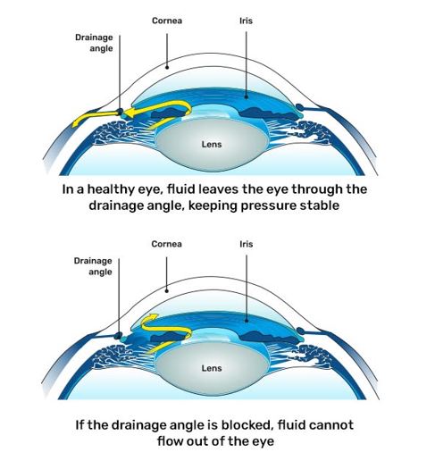
Our eyes constantly produce a clear fluid called aqueous humor which bathes and nourishes different regions of the eye. Typically, this fluid drains out of the eye through a drainage canal located at the angle of the eye. This angle is the junction between the cornea and the iris. In glaucoma, this fluid does not drain out as freely as it should. This increases the pressure inside the eye called Intraocular Pressure (IOP).
The normal intraocular pressure is between 10-22 mm of Hg. Raised IOP damages the optic disc, which is the part of the optic nerve seen within the eye. However, glaucoma can sometimes occur with a normal IOP – due to an extremely fragile nerve or reduced blood supply to the optic nerve. Hence, it takes more than measuring IOP to diagnose glaucoma.
Risk factors for glaucoma
The chances of glaucoma increase with age. So, it is mandatory after the age of 40 to have a complete eye examination. In case you have any of these risk factors, then do not delay your eye examination:
Family history of glaucoma
Research shows that the risk of inheriting glaucoma increases if several relatives have the disease.
Users of steroid tablets, drops and ointments
Certain medications may increase eye pressure as a side effect.
High plus or minus power
Changes in the shape and structure of the eye due to magnified powers can increase the risk of glaucoma.
Diabetes
Diabetes can double your risk of glaucoma (as per the National Eye Institute).
High blood pressure
Higher or lower BP can affect the intraocular pressure of the eyes.
History of eye trauma
Blunt or chemical injury to the eye, severe eye infections, and inflammatory conditions can lead to glaucoma.
What are the symptoms of glaucoma?
Glaucoma does not cause symptoms in the early stages. If you experience any of these symptoms, visit an ophthalmologist without delay.
Babies born with whitish-coloured eyes or bigger than normal eyes with watering and increased sensitivity to light should be screened for congenital glaucoma.
Types of glaucoma
Primary open-angle glaucoma
Angle-closure glaucoma
Developmental glaucoma
Secondary glaucoma
Primary open-angle glaucoma
The part of the eye through which the fluid flows out is open, but the drainage pathway is defective due to resistance to fluid outflow
It develops gradually without any symptoms
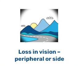
It affects the peripheral vision initially and, if undetected, progresses towards the centre
People are not aware they have the condition until they have disturbances in the central vision, which is the advanced stage of the disease
Angle-closure glaucoma
The drainage angle, which is part of the eye through which the fluid flows out, is narrow, due to which there is raised eye pressure
Gradual closing of the angle is called chronic angle closure, which behaves similarly to open-angle glaucoma
If the drainage angle closes suddenly, there is an acute rise in IOP, and it is called an acute angle-closure attack
The patient experiences pain, redness, nausea, vomiting, coloured haloes
It is an emergency condition and needs immediate treatment by an eye specialist
It generally occurs in people with high plus-powers
Developmental glaucoma
The third type, developmental glaucoma, is further divided into:
Congenital glaucoma, which occurs in infants from the time of birth
Juvenile glaucoma, which occurs in children and young adults
Secondary glaucoma
In certain types of glaucoma, there is an identifiable cause for the raised intraocular pressure and optic nerve damage
It may be caused by prolonged, indiscriminate use of steroids, severe diabetic retinopathy, eye injuries, inflammation of the eye (uveitis) or advanced cases of cataract
The part of the eye through which the fluid flows out is open, but the drainage pathway is defective due to resistance to fluid outflow
It develops gradually without any symptoms
It affects the peripheral vision initially and, if undetected, progresses towards the centre
People are not aware they have the condition until they have disturbances in the central vision, which is the advanced stage of the disease
The drainage angle, which is part of the eye through which the fluid flows out, is narrow, due to which there is raised eye pressure
Gradual closing of the angle is called chronic angle closure, which behaves similarly to open-angle glaucoma
If the drainage angle closes suddenly, there is an acute rise in IOP, and it is called an acute angle-closure attack
The patient experiences pain, redness, nausea, vomiting, coloured haloes
It is an emergency condition and needs immediate treatment by an eye specialist
It generally occurs in people with high plus-powers
The third type, developmental glaucoma, is further divided into:
Congenital glaucoma, which occurs in infants from the time of birth
Juvenile glaucoma, which occurs in children and young adults
In certain types of glaucoma, there is an identifiable cause for the raised intraocular pressure and optic nerve damage
It may be caused by prolonged, indiscriminate use of steroids, severe diabetic retinopathy, eye injuries, inflammation of the eye (uveitis) or advanced cases of cataract
Why choose the Glaucoma Clinic at KK Eye Institute?
The Glaucoma Clinic at K K Eye Institute is well-equipped to diagnose, treat and monitor glaucoma patients at all stages of the disease.
Top full-time Glaucoma Specialists with 20+ years of experience
Expertise in successfully managing complex glaucoma cases
Cutting-edge technology, equipment and treatment modalities
What are the tests needed to diagnose glaucoma?
Diagnosis of glaucoma requires a comprehensive eye examination and certain special investigations:
1. Slit lamp examination: This special microscope is the ophthalmologist’s stethoscope, and all patients suspected of having glaucoma undergo a slit lamp evaluation.
2. Applanation tonometry: The intraocular pressure is measured with an applanation tonometer attached to the slit lamp. This instrument is the gold standard for measuring the IOP. The non-contact tonometer uses an air puff to measure the IOP. It is good for screening but is not used for the diagnosis and treatment of glaucoma.
3. Gonioscopy: In this test, after numbing the eye, a contact lens with a mirror is placed on the cornea to visualise the area from where the fluid inside the eye drains out. By this test, we come to know whether the angle is open or narrow or if there are any pigments or tears in the angle.
4. Ophthalmoscopy: To diagnose glaucoma, we need to evaluate the optic nerve for signs of damage, and this is done by a dilated eye examination. A colour photo of the optic disc may also be taken for the documentation of optic disc damage and monitoring
If, in the above tests, you are diagnosed with glaucoma, or if there is a suspicion of glaucoma, certain specialised tests will be needed as follows:
1. Automated perimetry: This is a test to map the field of vision. It detects the visual field damage caused by glaucoma. One eye is tested at a time, and you would be asked to look at a central fixation: dull yellow light and peripheral white lights of varying intensity are shown. When you see the peripheral white light, you need to press a small button in your hand. This is an important test to detect the functional loss of vision due to glaucoma.
Also, it is repeated at regular intervals to know whether your glaucoma is stable or progressing.
The drawback is that if you move your eye too much or are inattentive during the test, it can cause errors, and the test may have to be repeated. So you need to be as comfortable as possible during the test.
2. Pachymetry: It detects the central corneal thickness. In this test, a small probe touches the central part of the cornea to measure the CCT. CCT influences intraocular pressure. Also, several studies have shown that those with a lower CCT have a greater risk of developing glaucoma and having progression as compared to those with a higher CCT.
3. OCT (Optical Coherence Tomography) Spectralis: This scan compares the thickness of the optic nerve head and the surrounding tissue (RNFL) in different quadrants with that of a healthy individual of the same age and can detect glaucoma in the early stage.
4. Anterior Segment OCT(ASOCT): This scan gives various measurements of the anterior chamber angle and can aid in the diagnosis of angle closure.
Treating Glaucoma - Saving Eyesight
Timely treatment of glaucoma is necessary to • Reduce eye pressure • Halt the progression of the disease to permanent blindness • Preserve the existing vision
Treatment is lifelong and requires regular follow-up. It depends on what type of glaucoma you have, and what treatments will work best for you. Your doctor will discuss the most suitable treatment for you after assessing your condition.
Treatment modalities can broadly be classified as:
Surgeons can easily switch between posterior and anterior laser treatment – a space-saving and efficient solution
It focuses the optimum amount of laser energy onto the point of treatment, offering sensitive, high-precision treatment to patients using a minimum of laser energy
The fine gradations permit optimum regulation of the laser energy for the sensitive, minimally invasive treatment of your patients
The high-grade laser beam source is fully integrated into the laser slit lamp
The compact format of the flexible positionable control panel offers handy control of the user-friendly interface
The unique 4-point aiming beam ensures a high degree of aiming accuracy
The symmetrical arrangement of the control elements makes all slit lamp functions ergonomically accessible
A 180° tiltable tube is used as a viewing device for the surgeon, allowing them to work
a
High-end Field Analyzer HFA-3-840
Reduce visual field-testing time with NEW SITA™ Faster, improving patient satisfaction with perimetric testing and reducing patient fatigue
Reduces setup time with a single trial lens
SmartTouch interface on the HFA3 platform gets you up and running with fewer touches
Kinetic perimetry advancements provide an easy-to-use graphical user interface with a full 180° testing range
Improved confidence in test results with RelEYE™ - Instantly review the patient’s eye position, at any stimulus point
Unsurpassed in efficiency, the system is patient-responsive:
It learns to perform as fast as the patient wants to go
Excellent surgical outcome: Microscope image with optimum contrast and detail recognition along with a large depth of field
A 1:6 ratio zoom system allows the magnification of the overall system to be set as required by the surgical procedures.
A 180° tiltable tube is used as a viewing
Schedule an appointment with our ophthalmologists today to protect your vision.
FAQS
As the incidence of glaucoma increases with age, it is mandatory after the age of 40 to have a complete eye examination. Risk factors include:
People with a family history of glaucoma
Those who use steroid tablets, drops or ointments
People with diabetes, hypertension or those who have had eye injuries
Those who have high minus or plus powers
People who see coloured rings around lights
It can also be seen in newborns, which is called congenital glaucoma, or in children and young adults, called developmental glaucoma.
Exercise: Research indicates that regular exercise can lower the risk of glaucoma. However, avoid lifting heavy weights, breath-holding asanas and head-down position yoga asanas such as sheershasana. They have been proven to cause an increase in eye pressure.
Diet: A diet rich in spinach, broccoli and sprouts, which contain lutein and zeaxanthin, helps avoid oxidative damage to the optic nerve. Avoid foods rich in trans fats, such as in deep-fried food, as they prevent the optimal functioning of omega-3 fatty acids and increase eye pressure.
Habit-forming Substances: Caffeine, alcohol and tobacco are known to increase eye pressure.
Musical Instruments: The trumpet and saxophone are known to cause an increase in eye pressure and are to be avoided.
Glaucoma affects the quality of life. A better understanding of your disease, treatment, and medications can make it easier to live with glaucoma and easier for your doctor to control your eye pressure. Early diagnosis, regular follow-ups and, therefore, early intervention are crucial in delaying the progression.
A complete ophthalmic evaluation is a must for everyone above the age of 40.
Get glaucoma screening done if you have a family history of glaucoma, diabetes, hypertension, heart disease, migraine, thyroid disease, history of eye trauma or if you are using high minus or plus power spectacles.
Babies born with whitish-coloured eyes or bigger than normal eyes with watering and increased sensitivity to light should be screened for congenital glaucoma.
Glaucoma is a lifelong disease which could be sight-threatening, but appropriate and timely diagnosis and management, regular use of medications and follow-up examinations can preserve the existing vision and minimise any further damage.
Regular follow-up for measuring eye pressure has to be done
Compliance with medications - Antiglaucoma medications have to be instilled according to the frequency prescribed by the doctor. Do not stop medications without consulting your doctor, even though you may not note a change in vision
Let your doctor know of any local side effects such as itching, swelling of eyelids, and redness developed after putting in the eye drops.
Follow-up tests, such as repeating visual field tests or OCT scans at intervals, as suggested by your doctor, to know whether glaucoma is progressing, must be done
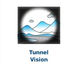
No, generally, there are no symptoms of glaucoma, such as redness or eye pain. In the early stages, there is no effect on the patient’s vision. As glaucoma progresses, the field of vision starts constricting, and in the advanced stages, only a small island of vision remains, and the patient can only see from one corner of the eye in the end stage disease, there is complete loss of vision.
The optic nerve damage caused by glaucoma cannot be reversed, but by appropriate treatment in the form of lowering the eye pressure by medical or surgical means, we can halt further damage and prevent blindness. So, though Glaucoma cannot be cured, it can be kept under control.
Generally, glaucoma does not cause redness or eye pain. In the condition called acute angle closure glaucoma, the drainage angle formed by the iris and cornea gets completely closed. The eye pressure rises in a short period in the range of 40-50 mm of Hg. The patient experiences redness, severe eye pain, blurred vision, and coloured haloes. This is a medical emergency and needs immediate treatment to lower the eye pressure and a laser iridotomy to open the drainage angle.
Glaucoma medications generally have to be continued lifelong. Patients should not stop the medications by themselves unless advised by the doctor.
Allergy may develop in some individuals due to the compound itself or the preservatives in it. There may be redness, itching, and swelling under the eyelids. The drop may have to be replaced with another drop from a different group, or preservative-free medication may have to be used. There is one class of drops known as beta-blockers, which are avoided in cardiac patients and patients with asthma as they affect the heart rate and lung bronchioles.
No, glaucoma surgery lowers eye pressure by creating an alternative pathway for fluid outflow and can halt the progression of glaucoma, thereby preserving the existing vision. But it cannot restore the vision which has been lost.